This article was last revised in 426 Days ago, some of its contents may have changed. If you have any questions, you can ask the author。
In the fascinating world of nature, lizards are renowned for their remarkable ability to change colors. These vibrant hues not only captivate our attention but also play a crucial role in the survival and reproduction of lizards. But what scientific principles underlie these dazzling colors? This article, in conjunction with the CIQTEK Field Emission Scanning Electron Microscope (SEM) product, aims to explore the mechanism behind the color-changing ability of lizards.
Section 1: Lizard Coloration Mechanism
1.1 Categories based on formation mechanisms: Pigmented Colors and Structural Colors
In nature, animal colors can be divided into two categories based on their formation mechanisms: Pigmented Colors and Structural Colors.
Pigmented Colors are produced by changes in the concentration of pigments and the additive effect of different colors, similar to the principle of "primary colors."
Structural Colors, on the other hand, are generated by the reflection of light from finely structured physiological components, resulting in different wavelengths of reflected light. The underlying principle for structural colors is primarily based on optical principles.
1.2 Structure of Lizard Scales: Microscopic Insights from SEM Imaging
The following images (Figures 1-4) depict the characterization of iridophores in lizard skin cells using CIQTEK SEM5000Pro-Field Emission Scanning Electron Microscope. Iridophores exhibit a structural arrangement similar to diffraction gratings, and we refer to these structures as crystalline plates. The crystalline plates can reflect and scatter light of different wavelengths.
Section 2: Environmental Influence on Color Change
2.1 Camouflage: Adapting to the Surroundings
Research has revealed that changes in the size, spacing, and angle of the crystalline plates in lizard iridophores can alter the wavelength of light scattered and reflected by their skin. This observation is of significant importance for studying the mechanisms behind color change in lizard skin.
2.2 High-Resolution Imaging: Characterizing lizard skin cells
Characterizing lizard skin cells using a Scanning Electron Microscope allows for a visual examination of the structural characteristics of crystalline plates in the skin, such as their size, length, and arrangement.
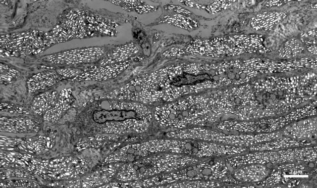
Figures1. ultrastructure of lizard skin/30 kV/STEM
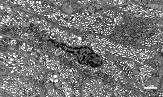
Figures2. ultrastructure of lizard skin/30 kV/STEM
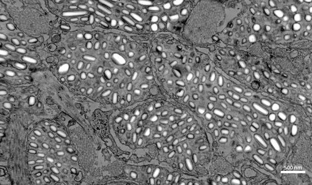
Figures3. ultrastructure of lizard skin/30 kV/STEM
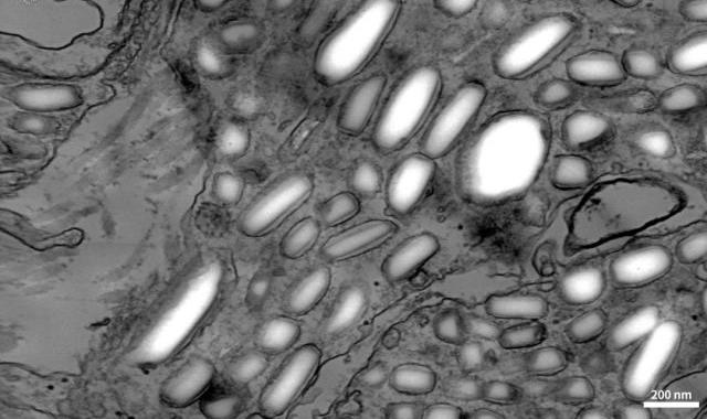
Figures4. ultrastructure of lizard skin/30 kV/STEM
Section 3: Advances in Lizard Coloration Research with CIQTEK Field Emission SEM
The "Automap" software developed by CIQTEK can be used to perform large-scale macro-structural characterization of lizard skin cells, with a maximum coverage of up to a centimeter scale. Thus, whether for high-resolution details or macroscopic area characterization, CIQTEK Electron Microscopes are capable of fulfilling the requirements.
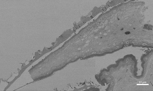
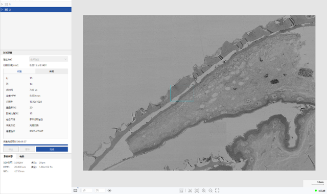
“Automap” Operation interface
CIQTEK Field Emission Scanning Electron Microscope (SEM) has the advantage of high-resolution imaging and supports the optional integration of a new type of Scanning Transmission Electron Microscopy (STEM) detector. It combines the features of both Scanning Electron Microscopy and Transmission Electron Microscopy to obtain high-resolution images formed by transmitted electrons at accelerating voltages of 30kV and below. It offers unique advantages for observing electron beam-sensitive biological samples.